Meluna Research is in the frontline of scientists working on biophotonics. Meluna stands for MEasuring LUminescence in NAture. From 2004-2007 the Meluna team worked closely with Prof. F.A. Popp, who was one of the first to study the behaviour of biophotonics. He hypothesized that the behaviour of biophotons provides an important indicator of the health and vitality of humans, animals and food. This knowledge is being further developed by the International Institute of Life Energy (IILE), among others. In IILE, most bio-photon pioneers are meeting regularly. Saskia Bosman was part of that team and she shares a lot more information on her website www.inspiradiance.nl
What are biophotons actually? How do you visualize their behaviour? What can this information mean for human, animal and plant health or for the health of the products we eat?
Living light
Bio-photons are photons in and of living systems. They play a role in the communication of cell-to-cell and organism-to-organism communication, in shaping the form field for biological development, in the coordination of metabolic processes, in the storage and transfer of energy. Moreover – as Popp (2007) and Van Wijk (2014) suggest – biophotons may play a role in our awareness and consciousness.

The discoverer of biophotones was Alexander Gurwitsch (explanation in the tekst below). At that time he spoke of mitogenetic radiation. Source: inspiradiance.nl
The Russian researcher Alexander Gurwitsch discovered, almost a century ago, that an onion root-point appeared to stimulate the cell division of another onion root at a distance of 2 mm. So ‘something’ had to be transferred between both plant roots. Later it was confirmed that there is indeed an interaction between the 2 plants when there is a tube of quartz around it. Nothing happened however with a tube of glass in between. The hypothesis was therefore that the phenomenon had something to do with UV-light. UV-light indeed passes through quartz and not through glass. But Gurwitsch didn’t have any UV measuring equipment yet. Moreover, after the Second World War, this kind of research stopped, because by then, molecular research emerged strongly and has supplanted further frequency research. In recent years however, this light research has celebrated its comeback.
Bio-luminescence.
Ultra-weak light also plays a role in the soil. In bioluminescence, ultraweak light is released in reactions involving oxygen radicals.
The production of light probably follows these chemical reactions:
The reduction process : NADH + H++ Luciferine –> NAD+ +LH2
The oxidation process : LH2+ O2 –> LO+ H2O + light.
Bioluminescence is functional in the communication between microbes: there are light-sensitive receptors in the cell membrane of many microorganisms, they stimulate each other’s cell division and they also regulate the spatial orientation and distance between the cells.
Particle or wave: what do we measure?
Do we see light as particles or as waves? That depends on how we want to measure them. With a photomultiplier as an instrument you measure particles, with a CCD-camera however, you measure waves. So your choice of the instrument determines how you measure light. Because the molecular approach in biology (particle-based) started to dominate after 1950, it became interesting for frequency researchers (wave-based) to look at the effects of biophotonics at the molecular level. After all, money was available for that research.

The photo-multiplier, an instrument that amplifies the extremely weak light signals in such a way that they can be seen on a photosensitive plate. Source: inspiradiance.nl
The photomultiplier was developed from the 1970s onwards for photon research considering photons as particles. It is called a multiplier, which means that more and more photons are released every next step. The most sensitive tubes release 1 electron per 4 photons. And so on, until enough electrons are released to be able to measure an electric signal: these signals together shape the image of light that we can see with our eyes on the photo paper or the computer screen.
From 1972 onwards F.A. Popp discovered that the photomultiplier tube is also suitable for photon research in living tissues. In West Germany, at the Internatonal Institute of Biophysics in Düsseldorf, he discovered that almost all life forms emit light in the visible and in the UV-domain. Apparently there is a biophotonic field around an organism. He also theorized that this photon field could play a role in the internal organisation of biological systems. This insight formed an important bridge between quantum physics and quantum biology. Since the 1970s, a third generation of biophotonics researchers has emerged: they look primarily at the information aspect of biophotonics, how is information transferred into a living system?
Highly light-sensitive CCD cameras are used when you consider light having a wave character. The measurement must be made in pitch darkness because the photon radiation is very weak. CCD cameras have also been improved and are capable now of counting individual photons. So you can count the number of photons on each pixel of the photo. Emission peaks can be recognized in the images: these are differences in the intensity of the photon emissions. For example, the skin underneath the nail stimulates the nail’s horn to emit more light. Something similar happens with a piece of white silk on the hand: the hand also stimulates the silk to emit photons. It may take hours before the total light pulse is processed into a still picture.

A very light-sensitive camera (above left) can register the very weak light impulses and make them visible to the eye. If you want to photograph a biophotonic image of your hand, you have to work in absolute darkness, put your hand under the camera and sit still and expose it for 30 minutes, the shutter opening time has to be 30 minutes to see something. Source: inspiradiance.nl
Studying the behaviour of photons, we should distinguish two types of emissions: Ultraweak Photon Emission UPE, and Delayed Luminescence DL. Both types of emissions are usefull as they give us specific signals of the metabolism and vitality of the tissue from which it is emitted.
- UPE (Ultraweak Photon Emission)
This is about excitation: electrons in an atom may jump into a wider orbit in which they can hold more energy. The molecule containing that atom absorbs that extra energy. Later on in the metabolism processes, the excited electron falls back into its smaller orbit and emits the energy difference between the two orbits in the form of a photon. The more the color shifts towards the UV, the shorter the wavelength and the higher the frequency, so the higher the energy. The further away the electron orbits are from the nucleus of the atom, the more energy is released when the electron falls back into a smaller orbit. You can measure the released energy with the CCD-camera which, however, takes minutes, sometimes hours. Photons emitted from a living system are photons that leak out of that system. The system cannot hold them, which provides information about the vitality of the system. The exact numbers of photons can be counted. We are talking about very low light intesitiy. This involves approximately 10 to 100 photons per second per square centimetre of tissue. This involves very little energy, varying between 10-18and 10-16Watts per square centimetre of tissue. Which is at least a thousand times lower intensity than the human eye can see. The colour – that means its frequency – of the ultraweak photon emission (UPE) provides information about the type of process from which photons are lost or about the element that emits them. For example, rapid cell division involves UV frequencies. If the colours are visible with the eye, then they refer to other reactions of biomolecules, such as proteins and fats reacting with reactive oxygen particles (Prasad, 2014).
- DL(Delayed Luminescence)
Delayed Luminescence – delayed release of light – happens in response to an external impulse of light, sound or heat. An organism stores light energy by means of excitation of electrons in biomolecules. This happens, for example, during photosynthesis in the chlorophyll of plants. Measuring DL is done with the photomultiplier tube. Leave the shutter open for about 10 seconds and measure the loss of photons during the 10 seconds. In the case of healthy living material or of crystals, this results in specific graphs with a hyperbolic decay curve (see the upper line in the graph below). This offers a clear distinction from non-living material or from diseased biological systems, producing exponential decay curves (the lower line). The figure below shows both curves. The exponential curve quickly decays to unmeasurable light intensity. Such a system does not have much internal coherence, it is sick or it is non-organic or non-crystal. The hyperbolic curve, on the other hand, decreases slower, which means that the photons are held longer, indicating a stronger internal order. And that is a sign of vitality of the measured product, which correlates well with its longer shelf life.

Delayed Luminescence of a product is measured during 10 seconds (horizontal axis). If your curve turns out to be a hyperbole, it indicates a healthy biological system or crystal. If the intensity (vertical axis) decreases faster, i.e. exponentially, this indicates a sick or a non-biological system. The figure shows for each curve a specific mathematical formula. The hyperbolic curve e–xand the exponential curve 1/x+1. This research allows to measure the vitality of food products, for example. In this case ‘vitality’ is an objective measure of the internal coherence and order in the product. Source: inspiradiance.nl
Hyperbolic versus exponential curves of Delayed Luminescence.
The hyperbole of a rotten tomato is no longer a clear hyperbole but comes close to an exponential curve. The decrease in light emission intensity is similar to that of dead material. A healthy tomato –in its ripening phase – gives a hyperbole, its structure is ordered, it is able to hold light longer and loose it later. You see this curve therefore above the exponential curve. It is striking that such a hyperbole curve is also found in crystals. This indicates a high internal coherence and ordered state of the molecules and atoms in crystals.
When a farmer treats his plants or animals with good intentions, the DL hyperbolic curve moves upward. In this way, good farming intentions give a different picture as compared to products grown without good intentions. This has become clear, for example, in research into the DL of organic milk and conventional milk. The measured results went against expectations: organic was not systematically better or worse than usual, while the intention of the farmer was the more important factor for the vitality of the product!
Are we sure about what we are actually measuring? Do we perhaps measure fluorescence? No, this is not the case, because fluorescence extinguishes long before luminescence begins. Do we perhaps measure light that is spontaneously emitted by the biological system? No, because we measure a impulse induced by an external source that was shining for only a short time on the tomato or the egg. The more the egg or tomato holds the photons, the slower the decay you measure in the DL. Does daylight also provide such an impulse? People do indeed store daylight: not only through the eyes, but also through the skin. Crystals can also store light and hold it more or less well. So living beings – and crystals – can be called light-holders.
How do biophotons occur?
The impulse from an energy source (in this case called ‘pump source’) gives more energy to an electron, which vibrates faster and – if it gets enough energy – jumps to an orbit further from the nucleaus. Sunlight – the most important pump source – excites electrons in chlorophyll in the leaves of plants. There are several other pump sources. Both in plants and in animals – and therefore also in humans – biomolecules can react with radicals of oxygen or nitrogen. In animals – that do not have photosynthesis – their food is a pump source. The most efficient light-producers are molecules with a ring shape including carbon and nitrogen, such as tryptophan (see picture in chapter 4.7).
Photons from a red lamp have a lower frequency with lower energy, so you can’t excite the electrons as much as with a blue lamp (that emits a higher frequency of light, so gives more energy). When the electron falls back into the orbit closer to the nucleus, it emits exactly this ‘excess’ energy again in the form of a photon. This energy can then be used, for example, for the metabolic process in the cell.
In summer we get more and more intense sunlight and then it is more blue, while in winter time it is more reddish. At midday the light is more blue than at the end of the day.
The much lower frequencies of audible sound can also act as a pump source and generate photon emissions. Red light can be compared to low tones and blue light to high tones. Loud sound can lead to light emission from an organism. You can see from its colour whether the photons come from oxygen or from another pump source. Normally, that oxygen is O2that is used for combustion, sometimes an oxygen atom is added (O3) or sometimes molecules are split up and free radicals are released. Interestingly, Aqua4D (see 2.3) advises music in pitch A when you need energy: that’s the tone of oxygen and it excites most. It is also the tone of the electron.

For measurements of ultraweak photon emissions from food, both spontaneous photon emissions and delayed luminescence are used.
The measurement of spontaneous photon emission is mainly used to get an impression of the plants’ immune system. Delayed luminescence provides information about the vitality of an organic product. Delayed luminescence is used to predict seed germination, estimate growth and ripening stages, and predict vitality under different storage conditions.
Here’s one type of computer graph showing the differences between healthy tomatoes (right-hand graph) and ‘normal’ supermarket tomatoes (left-hand graph). The healthy tomatoes conserve their energy levels.


Bio-photon behaviour pictured for supermarket tomatoes (at the left side) and very healthy and fresh tomatoes (at the right). The picture at the left shows that the tomato is leaking energy, wheres the picture at the right shows the tomato maintaining the energy and its order – it is more coherent. Source: Meluna.
I expect that these definitions of vitality will increasingly be accepted as a new quality marker of food products. It is complementary to commonly used tests for chemical content and structural properties of a product.
Biological functions of biophotons.
Biophotons play a role in cell-to-cell communication with the emission of ultraweak photons. A biological system – such as a cell – affects a neighbouring system with its photons. This type of communication not only takes place between cells, but has also been observed between organisms such as onion roots, radish seeds and water fleas. Although this has been known for almost a century, it is only in the last decades that this subject has regained the attention of scientists, as these phenomena can now be observed with modern sensitive devices.
The next question is, of course, how these photons can carry information. That’s in the patterns of their light waves. It is about wave-modulation. You can speak of light language, the way photons are ordered or broadcast in groups tells something about the process they come from.

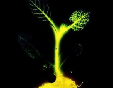
How do biophotons carry information? The hypothesis is that vibrations may carry specific deformations – called ‘modulations’ – specific for specific information. The picture above shows phase-modulation (Popp&Klimek 2007). The lower picture shows how ‘light-language’looks like: one counts emitted photons in time-lines. Source: inspiradiance.nl
And how is this information being transmitted in living tissue? Are there perhaps ‘cables’ that conduct the information-carrying biophotons? Such conductors have indeed been detected: in collagen tissue (Van Wijk, 2014). Proteins are deposited in collagen fibres, to which water molecules adhere. Water causes collagen fibres to swell. Water conducts light very well, so in this way light can spread through the organism. For example, you get communication from organs in the body to the pineal gland. For good internal communication it is therefore important to drink enough water.
Biophotones, delayed luminescence and the health of plants or eggs
Many different phenomena have specific effects on the health of a plant and we can make these effects visible by measuring biophotonic emissions. This emission almost always is related to the degree of (oxidative) stress in the plant.
If you put a plant in white light, within minutes you will measure the specific colour spectrum of active photosynthesis. 89% of spontaneously emitted photons of leaves have a wavelength between 600 and 1000 nm (red and near-infrared). It comes from chlorophyll and mitochondria in the cells. As soon as pathogens enter the leaf, one finds a different colour spectrum. Wounds or oxidative stress (O2, ROS, H2O2) induce higher photon emission. Any immune reaction in plant or animal creates an oxidating peak, visible in photon emission.
Measurements of biophoton behaviour of a plant can show stressy conditions before human eyes can see any stress in the plant. This early detectionby photon measurement offers the agricultural sector an opportunity to save money on pesticides. The earlier you see stress or illness coming, the less resources you need for control.
Beloussov, grandson of Alexander Gurwitsch, analysed the DL of chicken eggs to know whether or not they were fertile. He measured the changes over a period of 9 days. The yolk gives some light, the egg white does not. Cell biologist dr. Roel van Wijk has documented these issues in his great book: Light in shaping life (2014).
A non-incubated egg: its DL egg = DL of its scale.
An incubated egg, after one day: DL egg > DL scale + DL yolk.
The incubated egg, after 2 days: DL egg > DL scale.
After 9 days: DL egg < DL scale.
Beloussov’s explanation (2007) is that the embryo stimulates photon-emission of its scale during the first 2 days. While at day 9 the embryo probably ‘sucks’ photons from its scale.
At the moment, protein thickness is taken as a parameter of egg-quality. The quality of eggs must also be reflected in the electron flow and the photon flow in the egg. This is in line with the electrical measurements in (milliVolt and redox potential, etc.). It indeed is interesting to compare redox measurements with photon measurements The Louis Bolk Institute has already done a quality comparison of apples by means of structure, bio-photon measurements, Bovis values, chroma’s and redox potentials. http://www.louisbolk.org/downloads/1341.pdf.
Does genetic modification give a special picture? Indeed, one sees a DL curve of an exponential nature (as almost dead matter). This means that the system is internally less ordered, which in turn means that the organism is less adapted to the environment and has a lower self-regeneration potential.
In summary
We may state that this biophotonic concept of food quality and health, and its link with energy and coherence, in principle, is quite well in line with Schrödinger’s suggestion that life sucks in order, as expressed in his book What is Life (1944).
Food is used to maintain the state of life of the organism. In this way, living matter tries to avoid a relapse into the state of a thermodynamic equilibrium of dead matter. By avoiding the rapid relapse into the inert equilibrium state, an organism remains alive and healthy. For a long time we thought to feed on energy for its calorific value. Only later on one emphasised the concept of order. Organisms maintain themselves by continuously ‘pulling’ order from their surroundings, not only energy. This ‘order’ fits well in the MEI-picture, it could well be an aspect of Information that plants require in addition to Energy and Mass.